Contents
Microscopes are instruments designed to produce magnified visual or photographic images of objects too small to be seen with the naked eye. To achieve this, a microscope must accomplish three tasks: produce a magnified image of the specimen, separate the details in the image, and render the details visible to the human eye or a camera.
What Is a Microscope?
The word “microscope” is derived from the Greek words mikrós, meaning “small,” and skopeîn, meaning “to look at.” Literally, it is a machine for looking at small things. A more precise definition is an instrument used to see objects that are too small to be clearly seen by the human eye.
While our sense of sight is powerful, its resolution is limited. The average human eye can distinguish objects that are about 0.1 millimeters apart. The microscope opens up a world far beyond that physical limit. An optical (light-based) microscope is, in its simplest form, an optical instrument that presents a magnified view of an object. Using a microscope, one can observe the anatomy of organisms like insects, the fine crystalline structure of rocks, or the details of a single cell. The global microscope market was valued at USD 10.5 billion in 2022 and continues to grow as its applications expand. (Source: Grand View Research).
Who Invented the Microscope?
The invention of the simple microscope is often attributed to Antonie van Leeuwenhoek (1632-1723), a Dutch merchant with little formal education. However, the microscope’s creation was an evolutionary process. Around 1590, two Dutch eyeglass makers, Zaccharias Janssen and his father Hans, placed several lenses in a tube and discovered that an object at the far end appeared significantly magnified. They had invented the first compound microscope (an instrument with two or more lenses), though Leeuwenhoek would later perfect the single-lens design.
Leeuwenhoek crafted over 500 microscopes during his lifetime, which he reportedly never loaned to anyone. Through his meticulously ground lenses, capable of up to 270x magnification, he was the first human to see bacteria (which he called “animalcules”), red blood cells, and the life cycles of insects. For these pioneering discoveries, he is widely known as the “Father of Microbiology.”
Parts of a Microscope
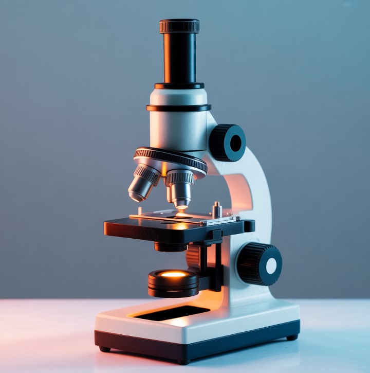
The primary parts of a standard compound light microscope are:
- Eyepiece (Ocular Lens): The lens you look through, typically providing 10x or 15x magnification.
- Stage: The flat platform where the slide (containing the specimen) is placed.
- Focus Knobs: The coarse focus knob makes large adjustments to bring the image into view, while the fine focus knob makes small adjustments for sharp detail.
- Condenser: A lens system under the stage that focuses the light source onto the specimen.
- Objective Lenses: These are the primary lenses on a rotating turret above the specimen. Common magnifications are 4x, 10x, 40x, and 100x (oil immersion).
How to Use a Microscope
First, you must prepare what you want to view, a process called mounting. To mount a specimen, place it on a glass slide and add a coverslip (a very thin piece of glass) on top. A drop of water or a staining agent is often placed between them to improve clarity. A great first specimen is a drop of pond water.
- Turn on the illuminator (light source) and ensure light is visible through the eyepiece.
- Place your prepared slide on the stage. Rotate the turret to position the lowest power objective lens (usually 4x) over the specimen.
- While looking from the side, use the coarse focus knob to move the lens as close to the slide as possible without touching it.
- Now, look through the eyepiece. Turn the coarse focus knob in the opposite direction, moving the lens away from the slide until the image comes into view.
- Use the fine focus knob to achieve a sharp, clear image.
To increase magnification, rotate the turret to the next objective lens and refocus using only the fine focus knob.
A Personal Experience: I remember my first time using a microscope in 9th-grade biology. We looked at a drop of pond water, and I was shocked to see a paramecium whizzing across the field of view, its cilia beating rhythmically. It felt like discovering a new universe. That single moment made biology tangible and real for me.
Types of Microscopes
- Simple Microscope: Uses a single lens for magnification, such as a magnifying glass, which typically provides up to 10x magnification.
- Compound Microscope: Uses a system of two or more lenses for much higher magnification. Most educational and laboratory microscopes are compound light microscopes, which use visible light to illuminate the specimen. Their maximum resolution is limited to about 200 nanometers by the properties of light itself. (Source: ZEISS Microscopy).
- Digital Microscope: These microscopes connect to a computer, often via USB, displaying the image directly on a screen. This allows for easy capture of images and videos, which can be saved as files. The market for these convenient tools is projected to surpass USD 400 million by 2028. (Source: Market Research Future).
- Electron Microscope: Instead of light, these instruments use a beam of electrons. A Transmission Electron Microscope (TEM) can magnify objects up to 50 million times, allowing scientists to see inside cells. A Scanning Electron Microscope (SEM) creates detailed 3D images of surfaces, resolving features as small as 1 nanometer. (Source: National Institute of General Medical Sciences, NIH).
- Scanning Probe Microscope (SPM): This class of microscopes uses a physical probe to scan a specimen’s surface, enabling the imaging of individual atoms.
Answering Your Questions
On social media, new microscope users often have questions. Here’s a common one:
A user on Reddit’s r/microscopy asks, “I just got my first microscope. What are some easy and interesting things to look at besides pond water?”
This is a great question. Start with household items. A single grain of sugar or salt under 40x magnification looks like a crystal palace. Other fascinating subjects include a strand of your own hair, the woven fibers in a piece of fabric, a thin slice of onion skin (a classic for viewing plant cells), or the intricate wing of a housefly.
The Microscope in the 21st Century
Today, microscopes are indispensable tools across nearly every scientific field.
- Medicine: Pathologists use microscopes to analyze tissue biopsies and identify cancerous cells. This is a critical step in diagnosing the nearly 1.9 million new cases of cancer detected in the U.S. each year. (Source: American Cancer Society).
- Materials Science: In the semiconductor industry, microscopes are essential for quality control, inspecting silicon wafers for defects on circuits with features smaller than 5 nanometers.
- Forensics: Scientists use comparison microscopes to match the unique striations on bullets, linking evidence to a specific firearm with a high degree of certainty.
- Modern Research: Advanced techniques like fluorescence microscopy, which uses fluorescent dyes that bind to specific molecules, allow biologists to watch cellular processes happen in real-time. As seen in a post by @NikonInst on X (formerly Twitter), showcasing a 2023 Nikon Small World in Motion winner, researchers captured the “neural dance” of a zebrafish’s nervous system, revealing stunning biological complexity.
Recommendations
- Getting Started: For hobbyists, a compound binocular microscope with magnification between 40x and 400x is an excellent and affordable choice. Brands like AmScope and OMAX offer popular entry-level models starting around $150.
- Virtual Practice: If you don’t have access to a physical instrument, you can practice using a virtual microscope. The University of Delaware offers a comprehensive online simulation perfect for learning the controls.


