Contents
The Amazing Discovery of X-rays!
Because he didn’t know what they were or what caused them, their precise nature remained a mystery at first.
‘X’ always stands for the unknown in math and science, so he picked ‘X-rays’ as a provisional name, a designation that stuck due to its aptness.
Because of this amazing find, he won the very first Nobel Prize in Physics in 1901, awarded “in recognition of the extraordinary services he has rendered by the discovery of the remarkable rays subsequently named after him.”
This news quickly got a lot of people excited, and many scientists started looking into the properties and potential applications of these mysterious new rays, recognizing their profound implications for medicine and science.
By January 1896, doctors were already using X-rays to look at bones and, later, lungs and other internal structures, revolutionizing medical diagnostics by providing an unprecedented view inside the living human body.
This was the start of radiology—the study of using X-rays and other forms of radiation for medical imaging to diagnose diseases and, eventually, for therapeutic purposes.
Let’s dive into the fascinating story of how he found these penetrating rays, a discovery that would fundamentally change medicine and physics.
The Discovery
You know about fluorescent light tubes, those long, cylindrical lamps that emit light when electricity excites a gas inside, causing it to glow. Back in the mid-1800s, scientists were studying what happened if you took a glass tube, sucked all the air out, and then put high-voltage electricity across electrodes at each end, creating a vacuum tube.
One end was called the ‘anode’ (positive side) and the other the ‘cathode’ (the negative side, from which electrons are emitted). When they did this, tiny particles (later called electrons) shot from the cathode to the anode at high speeds. As they traveled, these electrons hit the glass walls, making them fluoresce with a greenish-yellow glow, a phenomenon that intrigued researchers.
Scientists realized this glow came from ‘something’ that shot out of the cathode, went through the tube, and hit the glass walls, causing the visible light emission. They called this ‘something’ ‘cathode rays’ because it came from the cathode, the negative electrode in the vacuum tube.
The whole setup was called a ‘cathode ray tube,’ often specifically a Crookes tube, after the English physicist William Crookes, who developed improved versions. Today, we know that ‘something’ is a stream of electrons, fundamental subatomic particles with a negative charge.
This discovery was the first step towards finding X-rays, as the interaction of these cathode rays with other materials proved to be the key. Here’s how Roentgen stumbled upon his groundbreaking discovery.
Around that time, Wilhelm Roentgen, the German physics professor, started looking into what happened when these cathode rays, also known as electron beams, interacted with different materials outside the sealed tube. He wanted to see if cathode rays could escape the glass walls of the discharge tube and travel into the surrounding air. Roentgen covered his tube with thick black cardboard to block the cathode rays and turned it on.
Guess what happened?
He noticed a piece of cardboard covered with a special chemical (barium platinocyanide), a fluorescent material, that was lying on a table a short distance away started to glow with a faint but unmistakable greenish light. He turned the tube off and on, and each time, the glow on the distant screen synchronously appeared and disappeared, confirming its direct link to the tube’s operation.
This happened even though the tube was covered and the room was completely darkened, meaning no visible light from the tube itself could be reaching the screen. It was clear that some kind of rays were going right through the cardboard, the tube, and the air to reach that barium platinocyanide screen, causing it to fluoresce.
This amazed him, and he spent the next seven weeks researching this strange new form of penetrating radiation in almost complete secrecy, often working late into the night. He put the glowing paper further away from the tube, but the mysterious glow still persisted, albeit becoming fainter with increased distance, indicating the rays traveled a considerable range.
He thought maybe these were super-strong cathode rays or even a brand-new type of electromagnetic radiation, distinct from anything previously known. He already knew these rays could go through thick black cardboard, the glass of the tube, and the surrounding air, which was a remarkable property.
His next questions were: “What if I put something else between the tube and the fluorescent screen? How would these mysterious rays interact with different materials?”
Can these rays go through other stuff, just like they go through the opaque cardboard covering the cathode ray tube?”
So, he got to work and placed different, heavier materials between the tube and the barium platinocyanide screen to test the penetrating power of these unknown rays.
First, he used thin sheets of various materials like paper, wood, and aluminum. The paper still glowed, but not as brightly as before, indicating some attenuation of the rays. Then he tried heavier materials, including thin sheets of copper, silver, and even gold. He saw that some of the rays were blocked, but only platinum and lead could stop a significant portion of them, demonstrating their remarkable penetrating power through denser substances.
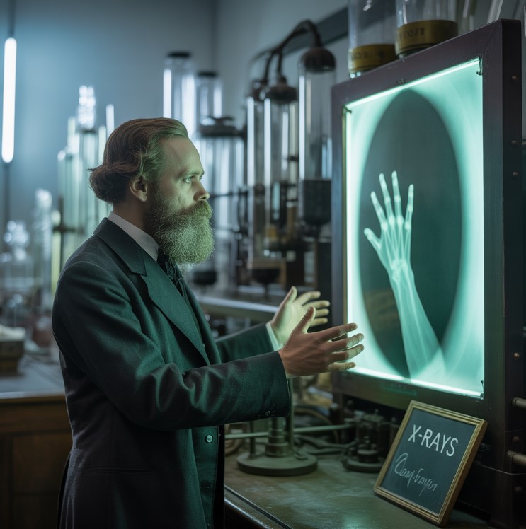
Roentgen’s next brilliant idea was to put his own hand in front of the path of the rays, between the discharge tube and the fluorescent screen. He was astonished to see that the rays went straight through his skin and muscle, projecting the shadow of his bones clearly outlined against the glowing background on the screen. From that moment on, medicine was forever changed, as a new era of diagnostic imaging had just begun. He had found a way to see inside things, even the living human body, without invasive procedures.
The inner parts of the body could now be seen without needing to cut into a patient, offering an unprecedented diagnostic tool. Roentgen loved photography and understood its potential for documenting scientific observations. Since photographic plates were already used in cathode ray research, it wasn’t hard for him to swap the glowing paper screen with a photographic plate to capture a permanent image.
This gave him a permanent record (a photo) of what he saw, which we now call an X-ray image or a radiograph. On December 22, 1895, he took the very first human X-ray picture, using his wife’s hand, complete with the clear outline of her wedding ring, a poignant detail that made the image even more impactful.
You could clearly see all the bones in her hand and even her engagement ring, starkly visible as a dense shadow against the transparent bone structure. It’s said that his wife, like many people at the time, felt a mix of wonder and profound unease, almost a sense of violation, at seeing her own skeletal structure.
Seeing her own bones made her feel strangely unsettled, remarking, “I have seen my death.” Because these rays were such a mystery, Roentgen called them ‘X-rays’—just like ‘X’ in math stands for the unknown variable or quantity. Mr. Roentgen never tried to patent his discovery or make money from its commercial applications, believing it belonged to humanity.
For ethical reasons, he gave his discovery to humanity so everyone could freely use and benefit from this revolutionary technology. He did receive the first Nobel Prize in Physics in 1901, but he donated all the prize money to his university, the University of Würzburg, for scientific research. Some say he even died quite poor because of World War I and the subsequent hyperinflation in the Weimar Republic, which significantly devalued his savings.
After the Discovery
News of Roentgen’s discovery spread like wildfire across scientific communities and the general public worldwide, captivating imaginations. Scientists everywhere could easily repeat his experiment because cathode ray tubes (like Crookes tubes) were already common equipment in physics laboratories, making replication straightforward.
By early 1896, X-rays were already being used in hospitals in the United States to look at broken bones and locate foreign objects embedded within the body, such as bullets or needles. In 1896, the Glasgow Royal Infirmary opened one of the world’s first X-ray departments, recognizing the immense medical potential of Roentgen’s discovery.
The head of that department, Dr. John Macintyre, took many impressive X-rays, including the first one of a kidney stone, a penny stuck in a child’s throat, and even a picture of the human head and brain (post-mortem), showcasing the diagnostic range. That same year, Dr. Hall-Edwards used an X-ray to diagnose a problem, finding a needle stuck in a woman’s hand, which he successfully located and helped surgeons remove.
For the next 20 years, X-rays were crucial in World War I, helping doctors find broken bones and shrapnel in wounded soldiers, significantly improving battlefield medical care and surgery. Eventually, people realized that too much exposure to X-rays could be harmful, leading to radiation burns, hair loss, and even increased cancer risk, prompting the development of safety protocols.
That’s why today we have special safety measures to protect both patients and medical staff, including lead shielding, controlled exposure times, and dosimeters. But even the harmful side of X-rays was put to good use, as their ability to damage cells was harnessed for therapeutic purposes.
In the early 1900s, they were found to be powerful in fighting cancer and skin diseases, leading to ‘radiotherapy,’ or radiation therapy, a specialized field utilizing ionizing radiation to treat malignant diseases. Radiology quickly became an important part of modern medicine, evolving into a cornerstone for diagnosis and treatment across countless medical specialties. As it grew as a diagnostic tool, it became its own specialized field with its own training, professional organizations, and a rapidly expanding array of imaging modalities beyond just X-rays.
At first, a radiologist did everything: they were the doctor, assistant, photographer, mechanic, and record-keeper—an impressive range of responsibilities for a single individual. But after 1900, these many tasks led to new jobs: the radiologist (the medical specialist), the X-ray nurse, the radiology photographer, and the radiology technician, each focusing on specific aspects of the imaging process.
Next, we’ll look at some interesting facts about Roentgen’s life, but first, let’s read a famous interview with him from Pearson’s Magazine published in April 1896, which provided an early public account of his momentous discovery.
Roentgen’s Own Words (Interview)
When did this happen?
“November 8th.”
And what did you discover?
“I was using a special glass tube covered in thick black cardboard, an opaque shield to block all visible light, experimenting with cathode rays. A piece of paper with a special chemical coating of barium platinocyanide was lying on a bench a few feet away. I was running electricity through the tube and saw a strange, faint light on the barium platinocyanide screen across the room, which immediately caught my attention.”
What was strange about it?
“It looked like something only light could illuminate or produce a fluorescent effect, yet no light source was apparent. But no light could escape the tube because the cardboard blocked all known light, even a strong electric arc lamp, making the observed glow profoundly perplexing.”
What did you think then?
“I didn’t just dismiss it as a minor anomaly; I immediately realized its potential significance. I figured the effect had to come from the highly evacuated Crookes tube itself, which was the only active apparatus.
I tested its penetrating properties by placing various objects between the tube and the glowing screen. Quickly, there was no doubt that these were entirely new and unknown rays. Rays were coming from the tube and making the distant barium platinocyanide screen fluoresce, even through opaque materials. I tried it from farther and farther away, even up to two meters (about 6.5 feet) from the tube, and the effect still persisted. It seemed like a new kind of radiation, unlike cathode rays, ultraviolet light, or any other known phenomenon. It was clearly something new, something no one had observed or systematically studied before, prompting my intense investigation.”
Is it light?
“No.”
Is it electricity?
“Not in any way we know.” “See how for him, almost everything about it was an enigma, a fundamental ‘X’ factor that he meticulously sought to unravel. That’s why he called them ‘X-rays,’ emphasizing their mysterious and undefined nature at the time of discovery.“
Fun Facts About Wilhelm Roentgen
- Wilhelm Roentgen’s parents, Friedrich and Charlotte, lived in a fancy house that was also their family textile business in Lennep, Prussia (now part of Remscheid, Germany).
- In 1848, during a revolution in Germany, his family moved to the Netherlands, supposedly to escape the political unrest and seek better opportunities, settling in Apeldoorn. So, Wilhelm spent most of his formative years in the Netherlands, attending local schools and developing a keen interest in nature. The plan was for him to take over the family business after finishing his education, as was common for sons in prosperous merchant families.
- He went to a technical school in Utrecht, but he actually got expelled in 1863 for supposedly drawing a rude cartoon of one of his teachers, though he denied being the artist. He claimed a classmate drew it, not him, but he refused to name the actual culprit, demonstrating a strong sense of loyalty. Because of this, he couldn’t get the certificate needed to formally enroll in a university in the Netherlands or Germany, a significant setback for his academic aspirations.
- While just sitting in on classes at Utrecht University, Roentgen heard from a friend that a new technical school in Zurich would accept students based on an entrance exam, even if they didn’t have the standard high school diploma, providing a lifeline for his education. He took the chance and started studying mechanical engineering at the Federal Polytechnic Institute in Zurich (now ETH Zurich) in 1865.
- After getting his degree in mechanical engineering, he switched to physics for his advanced studies and earned his PhD from the University of Zurich in 1869, under the supervision of August Kundt.
- Zurich was also where he met his future wife, Anna Bertha Ludwig, an event that profoundly shaped his personal life. He fell for Anna Bertha Ludwig, the daughter of a tavern keeper in Zurich, a woman known for her intelligence and warmth. They got engaged in 1869 with his parents’ blessing and married on January 19, 1872, beginning a long and devoted partnership. The wedding was a bit tricky because her parents, supposedly from a lower social class, weren’t entirely keen on their daughter marrying an academic, creating some initial social tension.
- In 1879, he became a full professor at the University of Giessen, where he continued his research into crystallography and electrodynamics.
- After winning the Nobel Prize, his fame also brought some unwanted scrutiny and public demands, disrupting his preference for quiet academic work. Arguments about who really discovered X-rays followed him for years, as several other scientists had experimented with cathode ray tubes and observed similar phenomena but had not fully recognized or investigated their significance. Since X-rays had always existed, and he was just the first to notice and study them, some other researchers felt they should have shared the recognition, particularly those who had seen early, fleeting evidence of the rays.
- Besides the Nobel Prize, he received many other awards for his discovery, like the Rumford Medal and the Matteucci Medal in 1896 and the Cresson Medal from the Franklin Institute in 1897, recognizing his immense scientific contribution.
- Wilhelm always preferred to work alone, immersed in his laboratory experiments, often shunning collaborators and assistants. He built much of his own equipment with great care and precision, showcasing his exceptional mechanical skills and attention to detail.
- His lectures weren’t popular with students because he didn’t give polished or particularly engaging presentations, focusing more on the experimental demonstrations than eloquent speeches. He even left a few notes about his discoveries and experiments and asked for all his scientific findings to be destroyed after his death, a request thankfully not fully honored, as some documents survived.
- Roentgen’s last years were filled with hardship and suffering due to the economic devastation of post-World War I Germany, including hyperinflation that wiped out his savings. His wife died after a long illness in 1919, and he retired from his post in Munich at the University of Munich in 1920, deeply affected by her loss.
- He spent much of his time at his country home near Munich, where he had a large garden and enjoyed pursuing his passion for hunting and nature. He continued to work and enjoy long walks in the countryside until the year of his passing, maintaining his intellectual curiosity and love for the outdoors. He passed away on February 10, 1923, from bowel carcinoma, a type of colon cancer, in Munich, Germany, leaving behind a monumental legacy.


